Scientists Show What Happens to the Lungs of COVID-19 Patients (Video)
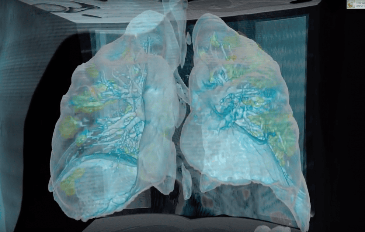
The video used the lungs of a 59-year-old man.
Doctors at George Washington University Hospital in the United States have created a 3D model showing what the lungs of a patient with the coronavirus COVID-19 look like. This is reported by CNN.
The yellow areas in the video indicate infected and inflamed parts of the lungs.
The scan shows that the damage is not localized to one area but instead covers massive sections of both lungs, showing how quickly the infection can spread, even in young patients. A patient with healthy lungs would have no yellow areas on the scan.
The video used the lungs of a 59-year-old man who is now in critical condition due to the effects of the coronavirus on his body. The images show extensive lung damage in an otherwise healthy man with high blood pressure.
Lung lesions at this stage of the disease are difficult to treat, and for 2-4% of patients with the new coronavirus, such damage can be irreversible.
The patient is at the stage of the disease where not only lung ventilation is required, but also another device that circulates the blood and saturates it with oxygen.
"This is not a 70- or 80-year-old patient, with immunodeficiency or diabetes. Apart from high blood pressure, he has no other significant medical problems," — explains Dr. Kot Mortman, head of the thoracic surgery department at George Washington University Hospital.
Dr. Mortman notes that young people can also fall into the risk group that currently includes older people. This is evidenced by the rise in hospitalizations of people of all ages.
Previously, it was reported that the United States may introduce a ban on resuscitation of patients infected with the coronavirus.
Map of coronavirus spread in Ukraine (updated in real time)
Zelensky: Russia Used 115 Missiles, Nearly 1,900 Guided Bombs and Over 3,000 Drones in a Week
Attack on Major DTEK Energy Facility in Northern Odesa Region Leaves Some Families Without Power

Britain Threatens Sanctions on Kremlin-Linked People; Poland Ready to Hand Ukraine Ammunition

Russia says it has completed "planned exercises" in the Black Sea

MVD: Fake Riots With Prop Blood Were Being Prepared Near Presidential Office

Law Enforcers Name Another Klitschko Deputy as Suspect

Berlin Holds Protest Against War in Ukraine; EU Prepares for Possible Russian Halt of Gas Supplies
North Korea Fires Ballistic Missile Over Record Distance in Five Years
Irish Fishermen Force Russia to Postpone Naval Drills

US sanctions in case of invasion of Ukraine could completely destroy Russia's economy, NYT reports